Hey Guys,
Since we are getting such a wide and varied case load of interesting tropical diseases, I thought it would be really cool if I did a whole blog devoted to some of the animals we have been treating. If you are either medically or bovine inclined (or both!), you would agree this is an interesting topic of conversation. If you are checking neither of these boxes, consider yourself warned and you may just want to wait for the next blog 😛

My lead off case is an animal with East Coast Fever (ECF) or Theileriosis. I start with this because it is probably to most significant disease of this region. It affects cattle, sheep and goats. The calf in the middle has it's head down and is depressed. In untreated cattle, the mortality rate reaches 100%, and even in treated animals the mortality rates are 20-40%.

A typical ECF animal will have a high fever (over 40 C), enlarged lymph nodes, pale mucous membranes, cough, and be very depressed. They may already be emaciated like this calf in the picture above.

Corneal edema is also a common finding, caused by infiltration of lymphocytes

This is the left pre-scapular lymph node, and it was one of the enlarged nodes.

Mucous membranes are pale, and sometimes there are linear red striations on the under surface of the tongue (not in this case).
ECF is caused by Theileria parva, which is a protozoal parasite, and it is transmitted by the brown ear tick, Rhipicephalus appendiculatus. Clinical disease is less common in the pure Ankole cows, the indigenous breed here. However, imported breeds are heavily affected by the disease.
The disease is found in Uganda, Kenya, Tanzania, Rwanda, Burundi, Malawi, Mozambique, southern Sudan,
Democratic Republic of Congo (DRC), Zambia and
Zimbabwe.
There are 28 million head of cattle at risk, and it kills one million annually.
Treatment involves injections of buparvaquone, given on day 1, day 3, and day 4 if necessary. Furosemide is added to most buparvaquone preparations, and injected parenterally together. The pulmonary edema associated with this disease can be severe, and can be the actual cause of death. Myocardial necrosis is also a factor. We also give dexamethasone daily to these cases, and fluids as deemed necessary.

Next up is babesiosis, another tick-borne disease. This 8 month old calf was acquired on market day, and became recumbent after making the walk home.

His temperature was 40.5 C, and there was a yellow tinge to his mucous membranes.

His lymph nodes were enlarged.
We didn't manage to snap a picture of it while he was urinating, but the urine was red-tinged. This is a big clue that you might be dealing with babesiosis, because this condition involves red blood cell hemolysis, causing the urine to be red with pigment. Another name for the disease is Redwater.

His ears were full of ticks. The same family of ticks (Rhipicephalus) are responsible for carrying the causative agents of babesiosis, which are also protozoan parasites. Babesia bovis and Babesia bigemina are the two main species responsible for disease.
We have been treating babesiosis with diminazine aceturate and oxytetracycline, as well as dexamethasone and an iron injection.

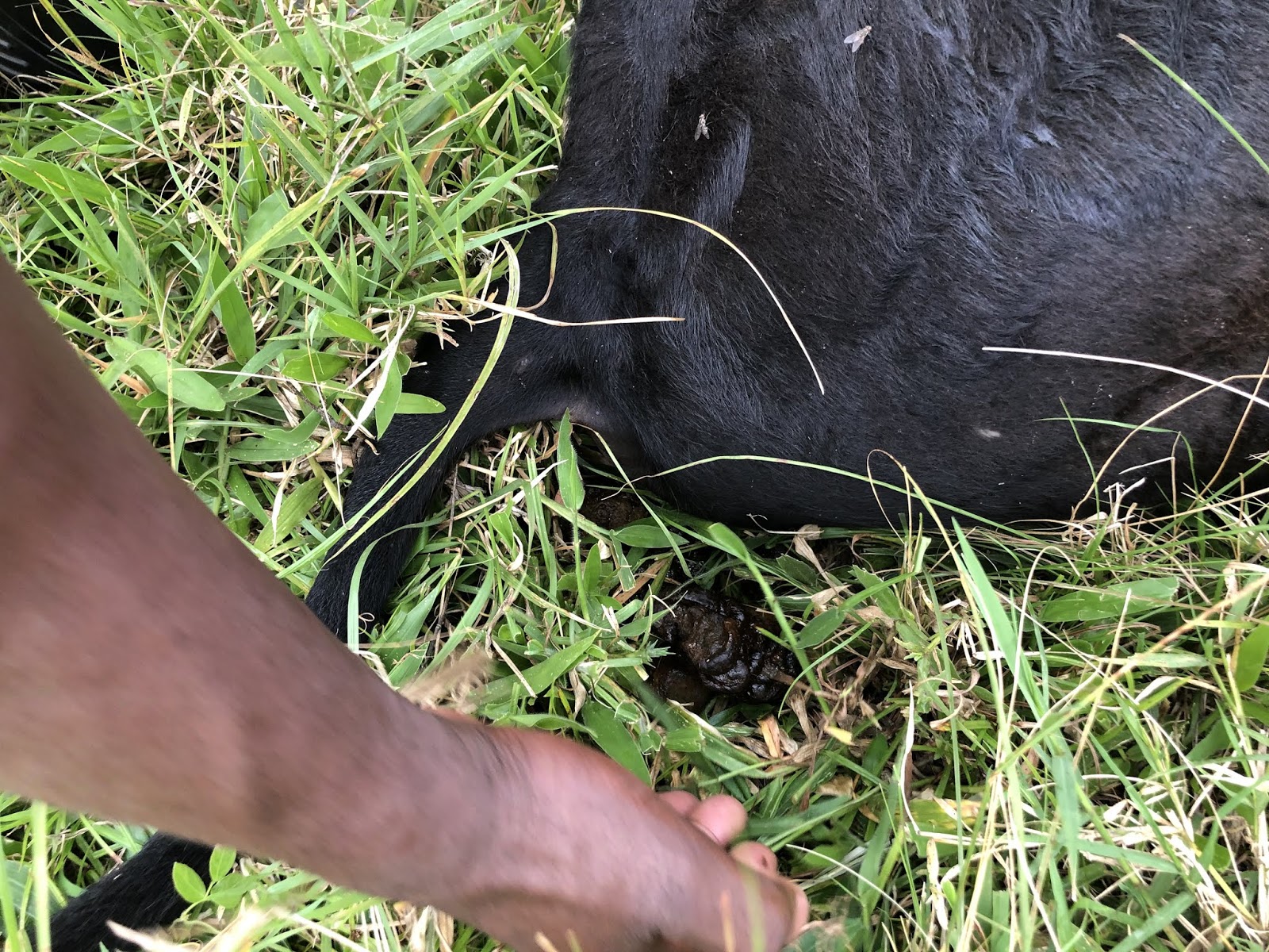
Let's round out the tick-borne illnesses with a case of Anaplasmosis in a mature milking cow. This cow calved in August, and has been waxing and waning clinically for the last month. As you can see from the picture, she was quite emaciated by this point. Kwesiga is taking a blood sample in this picture. Whenever farmers will give consent, he sends blood samples to the lab to corroborate his clinical suspicions. At the lab in Mbarara, they are making smears from EDTA blood to visualize the protozoal organisms inside the red or white blood cells. There are other (better) tests available for these three conditions I have mentioned so far, but they are not available at the closest lab and would likely be cost prohibitive for farmers here, anyways.
So what had us suspicious of anaplasmosis in this cow?
Apart from the ill thrift, she had:

Pale mucous membranes

She also didn't have enlarged lymph nodes, or a cough. This made us less suspicious of ECF.

That evening we started treatment with imidocarb and oxytetracycline. What is really interesting is that when we got the results back the next day, she was a strong positive for anaplasma and a weak positive for ECF. Cattle infected with ECF can become more susceptible to infection with other illnesses, especially the tick-borne ones. So in this case, this animal was likely first infected with ECF and cleared it. However, it did make her more susceptible to anaplasmosis, and this she succumbed to, causing the clinical signs. Her body was obviously putting up a pretty good fight, as her clinical signs were waxing and waning for over a month. Should the farmer have called sooner, sure. The good thing was that he agreed to testing, so when we got those results back we also instituted buparvaquone therapy, and she has since made a full recovery.

We were asked to evaluate this cow because she was slowing down and losing weight. This was not a tropical disease at all, but a great teachable moment. We have been teaching Kwesiga how to do rectal palpation for pregnancy diagnosis, and we are also helping him build up his physical exam skills. Earlier in the week we had been discussing all the things you could feel rectally (L kidney, rumen, pelvic floor, lymph nodes, etc). This cow had crepitus all around her L kidney, which can be consistent with pyelonephritis. With our limited supplies and diagnostics in the field, this would have to be enough to make a treatment plan for pyelonephritis. We asked the farmer to give her procaine penicillin for 5 days, and to re-evaluate her after that as it will likely need to be extended.

This is lumpy skin disease, caused by lumpy skin disease virus, which is in the Poxvirus family. It starts with fever, lacrimation, nasal discharge and hypersalivation, and then culminates with multiple eruptions on the skin. They are round, firm, raised and painful. What is worse for the animal is the fact that they are also in the mucosa of the GI, respiratory and genital tracts. What you see on the skin is just the tip of the iceberg. The condition is generally self-limiting and rarely fatal, but it carries a high production loss cost. It can be controlled with a modified live vaccine. The animal in the picture above has almost recovered from the disease.
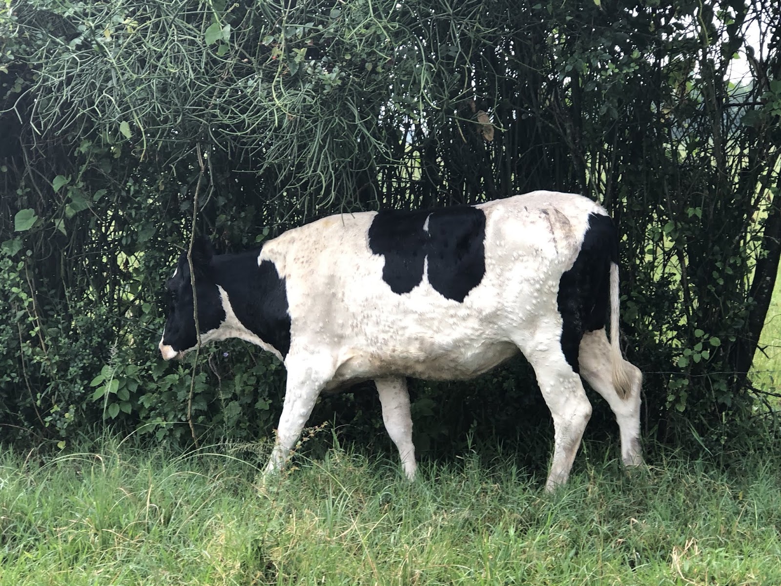
This animal is in the peak of her infection. Animals are usually viremic for less than 2 weeks, but the lumps may take months to fade.
I think I should end it there, because how much tropical disease fun can you handle in a single sitting?
Until Next Time!
No comments:
Post a Comment